Aktuellt
Blog Article – AI Meets Automation in Cell Microscopy
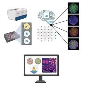
Smarter Imaging: AI Meets Automation in Cell Microscopy
Automation has already changed everyday microscopy by taking over tedious imaging steps and keeping conditions stable. Now AI is reshaping what we can learn from those images.
In this article, we show how automation and AI work together in high content imaging, combining consistent image sets from automated microscopes with AI based phenotypic readouts that detect subtle changes in cells. Together, they make experiments faster, more objective, and more reproducible.
We also look at how high content imaging is moving into 3D models such as spheroids and organoids, and how future AI guided microscopes could make imaging more predictive and easier to use.
For any questions, please contact:
Thommie Karlsson
Sales Manager Laboratory products
Mob: +46 73 800 95 75
thommie.karlsson@micromedic.se
Rickard Linnskog
Product Specialist Microscopy
Mob: +46 70 510 17 73
rickard.linnskog@micromedic.se
Johann Brännström
Product Specialist Microscopy LSR
Mob: +46 70 555 37 12
johan.brannstrom@micromedic.se